


The fungus of the nails (onychomycosis) in the legs is a disease that was developed as a result of the damage to the nail plates with mushrooms with dermatophytes (up to 96%), less often molds and yeasts (approximately 4%).The infection extends more frequently from the skin of the feet with long and existing mycosis.Here finds favorable conditions for development: greater humidity and nutrients.Under the influence of pathogens, the structure bothers and changes the color of nail plates.Over time, its complete destruction occurs.
Onicycosis is not only a cosmetic defect, but also a serious disease, which is subject to timely detection and adequate treatment under the supervision of a dermatologist.
The fungus in the legs is recorded in millions of people in the world.About 5% of the total population suffers from onychomycosis.The broader disease is common in people 50 to 60 years.Each second person is sick in this age group.The treatment of pathology is difficult for them due to the presence of somatic pathology, mainly vascular and endocrine.Men are sick more frequently than women.Older people get sick more frequently than young people.Children rarely suffer, mainly suffering serious diseases.With AIDS, the disease has an atypical image.
Causal agents of onychomycosis
The cause of legosis on the legs are different types of fungi: dermatophytes, yeast or mold -shaped fungi separately or in combinations.
- Dermatophyte fungi represent up to 90% of all onylomycosis.They are represented by the fungi of the genus Trichophyton (with most of the time T. Rubrum and T. Mentagrophytes var. Interdigitale).Most of the time, nail plates on the legs are affected by Trichophyton Rubrum.Dermatophytes are common in countries with a temperate climate.
- The fungus similar to the yeast of the genus Candida Onhomicosis in the legs rarely cause.They represent approximately 3% of all onylomycosis.In addition to Candida Albicans, fungi such as S. Tropicalis, S. Parapsilosis and S. Guilliosdii also cause the disease.
- Most mold fungi cannot cause nail fungi on their own.Only some of their species are independent pathogens: these are Scytalidium Hyalinum and S. Dimidiatum (Natrassia magniferese), which are not inferior in pathogenicity to dermatophytes.Leg onylomycosis is such molds as scopulariosis brevicaulis, aspergillus spp., Pyrenochaeta unguis-heominis, Alternaria spp., Fusarium spp.et al.The infection is more common in countries with a warm and humid climate: tropics and subtropics.
Epidemiology of the disease
Most of the onylomycosis are anthropophilic infection.They are sick and propagate the infection mainly to people.
Dermatophyte fungi
The deposit and the source of dermatophyte fungi are a sick person whose pathogens are transmitted with direct contact or their personal belongings.The infection almost always extends to the nails on the legs with the affected feet, whose disease proceeds both clear and secret (erased forms of mycosis).The risk of infection increases repeatedly in the presence of a disease in one of the family members.
The mushrooms are transmitted through infected shoes, clothing, files and scarves for nails, carpets, linen, towel, wipe, etc.The transmission of the infection occurs when the common bathroom, in the shower, sauna, pool, gyms and beaches.It contributes to the entry of fungi to the feet walking barefoot in common areas.Pathogens live for a long time in floors and wood floors.
Yeast -like fungi
The fungus similar to yeast of the genus Candida are saprophytic flora and always live in the skin of a person.An immune system is restricted by pathogens growth.The prolonged intake of antibiotics, contraceptives, glucocorticoids and cytostatic, endocrine pathology (often diabetes mellitus) and a series of diseases that exhaust the immune system.Explosive fungi penetrate skin nails and mucous membranes of the same patient, or enter the human body with infected products rich in carbohydrates.
Molds
The molds live on the ground.Their disputes fall into environmental products, things and objects.Nedimatophytes do not spread among people.

Risk factors for disease development
For fungi, dermatophytes are characterized by a hereditary predisposition, male sex, old age, vascular diseases, diabetes mellitus, states of immunodeficiency, greater sweating, nail injury and the presence of other dermatomycosis.
The infection of fungus similar to the yeast of the Candida genus is characterized by an increase in temperature and moisture, immunodeficiency states, increased blood glucose, nail injury and lack of complement with personal hygiene rules.
For mold infection, severe immunodeficiency states and nail injury are characteristics.
Groups at risk
The Risk Group in the development of ONYKHOMICISIS includes:
- People constantly wear changing rooms, showers, saunas, etc.
- Professional athletes (swimmers, soccer players, athletes, etc.).
- Military personnel and other groups of people who wear patented shoes.
- Male faces.
- Age is over 60 years.
Contribute to the development of the fungus in the legs:
- Using tight and well adjacent shoes.
- Increase in sweat or dry legs.
- Injuries and nail abrasions, feet scratches, incarnate nails, etc.
- Hot and hot climate accommodation.
- Walking barefoot in public places.
- The presence of skin diseases in which nail keratinization (psoriasis, ichthiosis) is interrupted.
- Diseases such as diabetes mellitus, immunodeficiency states, circulatory disorders of the lower extremities, blood disease, prolonged intake of corticosteroids, antibiotics and cytostatic.
- Genetic predisposition.
Fleet fungi development routes
There are several ways to penetrate fungi in the nail plate:
- Distal or distal-lateral (from the free or lateral edge).
- Superficial (directly through the nail plate).
- Proximal (subty -gut).
Distal-lateral path of fungi penetration
The route of the penetration of distal or distal lands is characteristic of Trichophyton Rubrum fungi.The pathogens are introduced into the nail plate from the free (distal) edge or the lateral regions (lateral edge).The main inflammatory process occurs in the bed of the nail, where there is a greater cell proliferation.The skin tacaño layer on the free edge is thick (hyperkeratosis), as a result of which the nail plate rises and exfolia (onicolysis).
In addition, the infection extends in the direction of the hole and penetrates the nail plate, which gradually (slowly) is destroyed.With the damage to the matrix, a total distribution onylomycosis occurs.
Hyperkeratosis of the nail bed is observed in chronic eczema, psoriasis, warts, plain red lichen.
Superficial path of fungi spread
Trichophyton Mentagrophytes Var.Interdigita is more aggressive in relation to the horny structures of nail plates than other dermatophytes.They mainly affect the outside of the nail plate, causing the development of white surface onicycosis.Fungi under the influence of keratinaz enzymes pierce the stratum layer with hyphae, gradually capturing all layers of the nail plate.Mainly 1 and 5 fingers are affected.They are they who are subject to the biggest shoes trauma when walking.In the disease, 1 and 4 intercal folds are affected.
It is believed that the superficial form of onychomycosis can also be caused by non-humanatophytes fungi: acremonium spp., Fusarium oxysporum and some types of Aspergillus.
Proximal fungal distribution route
There is a third path of mushroom penetration on a nail plate: through a proximal nail roller and a nail bed.The defeat begins with the skin in the nail roller area, which is thick and exfolia from the nail surface.In addition, the final part of the matrix and the bed of the nail is involved in the process, with damage to which furrows, irregularities and cracks appear in the nail.With the penetration of pathogens in the nail plate, the nail acquires a white opaque color over time.Over time, complete destruction and loss of the nail plate are observed.It is found more frequently in patients infected with HIV with the spread of infection through blood vessels.
Characteristics of the damage to the fungus similar to the yeast of the genus Candida
The damage to the fungi of the genus Candida begins with a Paronichia inflammation of the proximal roller (located near the hole).Its edema and thickening are observed, which leads to the separation of the cuticle from the plate surface.In addition, mushrooms fall freely in the matrix and the bed of the nail, causing the nail of the finger tissues over time.
Characteristics of damage to fungi by incorporators
The damage of the nails to the mushrooms with non -heartbeat is secondary.Molds (often Scytalidium spp.) Placed in the already affected cracks, space between the nail bedshaw or the desolate vessels.Next, hyperkeratosis develops and the slow destruction of the nail plate.
Clinical forms of legosis on the legs
There are several foricomycosis on the legs:
- Distal side.
- Superficial white.
- Proximal.
- Total distribution.
Distalaral submarine-lalateral onicycosis on the legs
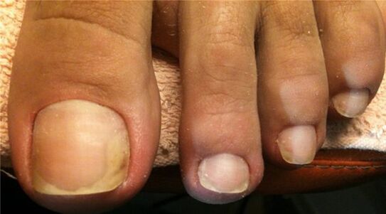
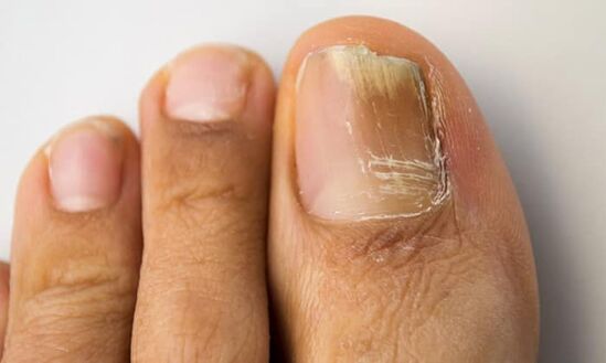
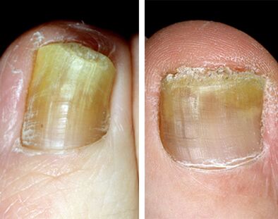
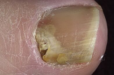
This form of disease is the most common.In most cases, the cause of onychomycosis is dermatomycetes, particularly Trichophyton Rubrum.The pathogens penetrate the nail plate from the side of the free edge and the side edges.Pereneophaeum hyperkeratosis is developed, as a result of which there is a detachment of the finger tissues (onicolysis), loses transparency, acquires a whitish or yellow color, begins to crumble.With the development of underwater hyperkeratosis, the nail plate seems thickened.With the progression of the disease, the focus of the lesion expands to the hole, as indicated by emerging yellow strips.Over time, the entire nail plate and matrix are involved in the pathological process, which eventually leads to dystrophy and destruction of the nail.
In elderly people, pronounced hyperkeratosis (thickening), onhogrifosis (thickening and deformation in the form of poultry claws) or coilonichia (concave deformation) is often observed.Your nails are often affected by mixed flora: dermatophytes, molds and even bacteria.
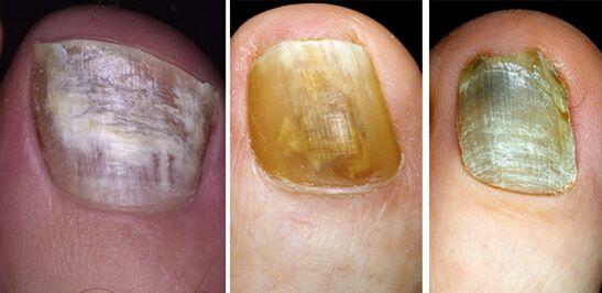
Superficial (white) form of onicycosis on the legs
Onylomycosis of the white surface in the legs is the second largest form of distribution damage.Its cause is mainly trichophyton Mentagrophytes var.Interdigitale, which penetrates the nail plate directly through its upper part (pre-system), as well as some types of fungi and hectophites.Mainly affected by the nail in the first finger of the leg, less frequently, the fifth.
At first, small white spots appear and strips on its surface, which eventually captivates an increasing surface.Little by little, the color becomes yellow, ocher.The nail surface becomes loose, rough, powdered, jumps easily.Thickening and separation from the bed of the nail do not occur.
The proximal underwater form of legosis on the legs
This form of mycosis is a rarity.It represents approximately 3% of all onicycosis.The reason is that the fungi of Candida Albicans and Trichophyton Rubrum by Candida Albicans.Nail candidiasis is preceded by the inflammation of the periodogue roller.He swells, acquires red, becomes bright.The cuticle rises and the infection penetrates the final part of the matrix and the bed of the nail, when damaged by the groove, irregularities and cracks are observed in the nail plate, loss of natural brilliance and clouds are observed.Little by little, the nail is destroyed, in severe cases it disappears.This form of legs in the legs is often found in HIV -infected patients.
Total distribution form of legosis on the legs
This form of onychomycosis develops more frequently with a long -term current (chronic course), whose cause is more often the fungi of Trichophyton Rubrum and Candida Albicans.At the same time, nail plate, bed and matrix are involved in the pathological process.Excuse the nail occurs as a result of the development of underwater hyperkeratosis.Over time, the nail plate is destroyed and the new one due to the affected matrix does not grow or grow badly.
Types of damage to nail plates
There are 3 options for onylomycosis:
- Normotorophic.
- Hypertrophic.
- Atrophic
Normotrophic type of legosis on the legs
With a normotrophic type, the infection is located in the upper layers of the nail plate.Its thickness and color in the disease do not change, but spots and stripes are visible in the depths.The color of the nails varies from white to saturated yellow.After a while, spots and stripes merge.The damage area extends to the entire nail plate, excluding the moon.It is not observed to break and lock.Sometimes there is a slight loosening of the free edge.With proper treatment, a cure is possible.
Hypertrophic type of legosis on the legs
This type of onylomycosis is the most common.As a result of the development of underwater hyperkeratosis, the nail plate is significantly greased, its brightness is deforms and loses.The nails become unequal, opaque, acquire a brownish gray color and crumble.The moon area is not affected.The disease gives the patient tangible discomfort.In elderly patients, the development of onhogrifosis is observed: nails are thickened, lengthened and bent like the claw of a bird.
Atrophic Type of Leg Onylomycosis
With an atrophic type (onicolithic), the nail plate quickly loses its connection from the nail bed, many gaps appear in its layers, it fades, it becomes thinner and changes color to whitish or yellow.The surface remains soft for a long time.Over time, partial destruction occurs.
Signs and symptoms of nail fungus
Most of the time, the change in the nail begins with a free (distal) or lateral (lateral) edge.
Color changeWith onychomycosis, a change in the color of the nail plate is the first sign of the disease.It becomes opaque, often loses its brightness, acquires a white or yellow color, with overlap with mold mushrooms: brown, brown, green and even black.
Thickening.The increase in the number of horny masses as a result of the development of underwater hyperkeratosis leads to the thickening of the nail.
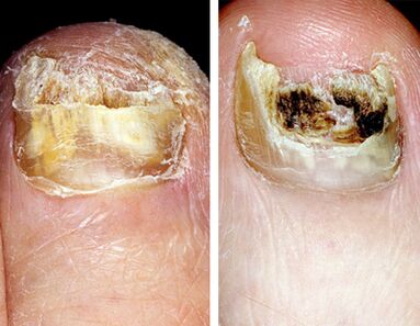
Overwhelming and destruction.In case of illness as a result of the vital activity of fungi, the nail plate first falls apart and, over time, over time, completely destroyed.
Characteristics of nail damage with different types of onylomycosis
Nail damage with different types of fungal diseases has its own characteristics.The main types of pathogens are Trichophyton Rubrum (70 - 90%) and Trichophyton Mentagrophytes V.Interdigitale (8-30%).Candida albicans, Moho fungi, T. Mentagrophytes V.Gypseum, T. Verrucosum, T. Tonsuras and T. Vioceum, Epidermophyton floccosum, Trichophyton are much less common.Schonleinii.
Rubrophic legosis onylomycosis
Rubrophic in the Russia Federation represents 70 to 90% of all mycosis.The feet in the disease are most frequently affected (usually a type of drying).An indispensable satellite of feet sform is a nail fungus on the legs.With mycosis, the distal-dilutal shape of onychomycosis usually develops, pronounced hyperkeratosis is characteristic, several fingers in the leg are affected at the same time and, often, the fingers by one hand.The disease proceeds without special subjective sensations.Pain and discomfort when wearing shoes occurs with pronounced hyperkeratosis, initiation and an embodied nail.The source of infection is often in the patient's family.
Often, associated onychomiosis are recorded: Trichophyton Rubrum and Candida Albicans, Trichophyton Rubrum and molds.It is important to evaluate the cultural study.
Leg onylomycosis with t.Mentagrophytes fungi.V.Interdigitale
Monores T. Mentagrophytes.V.Interdigitale affects the skin of the feet and nails.Epidermophtosis represents 10 to 30% of all mycosis of the feet.
With the disease, the upper (dorsal) of the nail plate is affected.The superficial white shape of onychomycosis usually develops.The pathological process is mainly involved in 1 and 5 fingers (they are subject to the largest trauma for shoes during shoes during the walk) and 1 and 4 folds between packages.The transmission of infection occurs when a common bath is used, in the shower, sauna, pool, on beaches and swimming pools.
Leg onylomycosis with damage to the fungus similar to the yeast of the genus Candida
This form of legosis in the legs is a rarity.It represents less than 3% of all onylomycosis.Often, the disease is recorded in people with chronic generalized candidiasis.The damage to the nails, as a rule, begins with the inflammation of the periodogue roller located near the hole.Its edema and thickening are observed, which leads to the separation of the cuticle from the plate surface.In addition, mushrooms fall freely in the matrix and nail bed (proximal sub-basic shape), if slots, irregularities and cracks appear in the nail, a loss of natural brilliance and clouds appear, a brown brown brown color is manifested.Little by little, the nail is destroyed, in severe cases it disappears.
Leg onylomycosis caused by mold
Plastic fungi are filled in an affected nail: cracks, in the spaces between the nail beds or desolate vessels.Next, hyperkeratosis and slow destruction of the nail plaque are developed, which during the disease is dyed black (Scytalidium spp.) Or green or gray (scopulariopsis brevicaulis).
Diagnosis of onychomycosis
The diagnosis of onychomycosis is based on data from epidemiological history, the clinical image of the disease and the data of the laboratory research method.
In a microscopic examination of the material, the nature of the disease (fungus or other pathogen) is established.Fungi identification is established with a microbiological examination (material crops in a nutrient medium) with the posterior microscopy of a pure crop.The process is laborious, success is achieved in half cases.The correct collection of affected nail material is the key to a successful microbiological study.
Differential diagnosis
Only in half of the cases of patients with dermatologists with changes in the shape and color of the nails make fungal diseases.Onychomycosis should be distinguished from eczema, psoriasis, Reitera's syndrome, Pachionichia, Daria's disease, plane lichen, Norwegian scabies, bacterial lesions.